

How To Clean Ortho K Lense With Boston Multi Action Solution
![]()
The Effect of Dissimilar Cleaning Methods on Protein Deposition and Optical Characteristics of Orthokeratology Lenses
1
Department of Chemical Engineering and Biotechnology, National Taipei University of Applied science, i, Sec. three, Zhongxiao East. Rd., Taipei 10608, Taiwan
2
Section of Ophthalmology, Chang Gung Memorial Hospital, Linkou, No. 5, Fuxing St., Taoyuan 333, Taiwan
3
College of Medicine, Chang Gung University, No. 259, Wenhua 1st Rd., Taoyuan 333, Taiwan
iv
Research and Development Center, Brighten Optix Co., 6F-1, No. 150, Sec. 4, Chengde Rd., Shilin Dist., Taipei 111, Taiwan
5
Department of Optometry, University of Kang Ning, No. 137, Alley 75, Sec. 3, Kang Ning Road, Neihu District, Taipei 11485, Taiwan
vi
Institute of Biomedical Engineering and Nanomedicine, National Health Research Institutes, No. 35, Keyan Road, Zhunan Town, Miaoli County 35053, Taiwan
*
Author to whom correspondence should exist addressed.
†
These authors contributed equally to this piece of work.
Academic Editors: Bramasta Nugraha and Faisal Raza
Received: 29 October 2021 / Revised: half-dozen Dec 2021 / Accustomed: 7 December 2021 / Published: nine December 2021
Abstract
Orthokeratology lenses are usually used for myopia command, especially in children. Tear lipids and proteins are immediately adsorbed when the lens is put on the cornea, and protein degradation may cause discomfort or infection. Therefore, we established an in vitro protein deposition analysis by mimicking the current cleaning methods for orthokeratology lens wearers for both brusque-term and long-term period. The results showed that the amounts of tear proteins accumulated daily and achieved a balance afterwards xiv days when the lens was rubbed to clean or non. Protein deposition also affected the optical characteristics of the lens regardless of cleaning methods. Our results provided an in vitro analysis for protein deposition on the lens, and they may provide a potential effective method for developing intendance solutions or methods that tin can more effectively remove tear components from orthokeratology lenses.
1. Introduction
Orthokeratology (ortho-grand) lenses take been used for affecting vision since 1945, when the corneal contact lens was fabricated with plastic [1]. Modernistic ortho-1000 lens tin can be worn overnight to reshape the anterior cornea by the designs of reverse geometry; thus, patients with myopia practise non need to wearable glasses for vision correction during the daytime [2]. The safety of wearing ortho-g lenses has been monitored by several long-term clinical follow-ups, and complications still arise, although the majority was not immediate agin events [3]. The major complications are microbial keratitis and superficial corneal staining, while microbial keratitis is a potentially vision-threating complexity [4]. The possible cause of microbial keratitis might due to lack of eye movements which unremarkably help to disrupt the bacterial glycocalyx, resulting in less spreading of lysozyme over the surface of eye tissue and making eyes being more than susceptible to the infection [5]. In addition, microbial keratitis might exist non-compliance, including inappropriate lens intendance and wearing continuously despite meaning discomfort [6,7]. Indeed, tear proteins and lipids are immediately deposited on the surface of the lens when a contact lens is put into the eye. Tear proteins are too easily accumulated on the lens and may cause immune reactions if lenses are not existence cleaned completely [viii,9].
Ortho-yard lenses can be reused for at least one year; thus, lens care is a more important and critical compared with soft contact lenses that are made equally disposable daily. It has been shown that cleaning rigid contact lenses with lens care solutions without finger rubbing was not an effective method for removing stubborn materials (such as mascara and hand cream) from the lens [10]. Rigid contact lens multipurpose intendance solutions, hydrogen peroxide, and povidone–iodine are all advised for cleaning ortho-g lenses, but the related studies but focused on disinfecting microorganisms [7]. The studies near removing tear components from ortho-k lenses have not yet been investigated intensively. In addition, it is also possible that an accumulation of tear components may touch on the opposite geometry of ortho-1000 lens, resulting in ineffectiveness in flattering the cornea. The relationship between tear components aggregating and the optical characteristics of ortho-k lenses is still unclear.
In this study, we first investigated protein deposition in the absence or presence of tear lipids. Two tear proteins were analyzed hither: the lysozyme and albumin. Lysozyme is the most arable tear poly peptide, while the concentration of albumin increases when the eye is closed or when wearing ortho-k lenses [11,12]. Protein adsorption amounts on ortho-g lenses were analyzed by cleaning with or without finger rubbing. Both short-term and long-term protein degradation analyses were conducted, and the accumulated protein amounts were quantified daily. In add-on, the optical characteristics of ortho-grand lens after a long-term protein deposition procedure were also investigated.
2. Materials and Methods
ii.ane. Orthokeratology Lenses, Contact Lens Care Solution, and Artificial Tear solution
The material of ortho-yard lenses used in the study was Boston XOTM, and the fabric generic name was Hexafocon A (Brighten Optix Co., Taipei, Taiwan). The contact lens intendance solution used for cleaning and rinsing was Menicon Intendance Plus multipurpose solution for all rigid gas permeable lenses (Menicon, Nagoya, Japan). The preparation of artificial tear solution has been published previously [13]. In general, a complex of salt solution was made according to the concentration listed in Table 1 [13]. The pH value of a complex of table salt solution was adjusted to vii.4 and was stored at room temperature for 3 or more than days. The 2000 times lipid stock solution was made in a solution of 1 hexane:ane ether, and the stock concentration was listed in Table 1. Then, 250 μL of 2000 times lipid stock solution was added into a complex of salt solution, and the mixed solution was placed into an ultra-sonic bath at 37 °C. The mixed solution was sonicated at 90 W and purged with nitrogen gas until the lipid stock solution was fully incorporated, and the olfactory property of hexane and ether was cleared out. Then, either lysozyme or albumin was added into the mixed solution containing salts and lipids for the further experiments. All the components of table salt, lipid, and poly peptide were purchased from Sigma-Aldeich (St. Louis, MO, United states).
2.two. Short-Term Protein Deposition Analysis
The ortho-k lens was treated by an oxygen plasma organization (Force State OP300, PFS Co., Ltd., Taipei, Taiwan) at 100 mTorr, 80 W, 10 standard cubic centimeter per infinitesimal (Sccm) for 120 south before the experiment, in society to ameliorate the hydrophobicity. The orthokeratology lens was placed in 2 mL of solution 1, which was artificial tear solution containing either 2.0 mg/mL lysozyme or 0.2 mg/mL albumin, and incubated at 37 °C for 8 h (Step 1 in Figure 1). Then, the immersed lens was placed in ii mL of care solution at 37 °C for sixteen h (Step ii in Figure one). Finally, the ortho-thou lens was not cleaned (no rubbing) by intendance solution and was placed into a new bogus tear solution. The other grouping was cleaned (rubbing), rinsed with fingers in gloves past 50 μL of care solution, and placed into a new artificial tear solution (Step 3 in Figure ane). A completed cycle (step ane to 3) was repeated 5 times (five days), and the protein concentrations in each solution were analyzed.
The Bio-Rad DC protein assay (Bio-Rad, Hercules, CA, USA) was used for measuring the corporeality of lysozyme or albumin in each solution [14]. The optical density (OD) value was obtained by an Enzyme-Linked Immunosorbent Assay (ELISA) reader with a wavelength of 280 nm. The deposited protein concentration on the orthokeratology lens afterward 3 steps on each was calculated as follows: (the original protein concentration)–(protein concentration in solution ane)–(protein concentration in care solution) for the no rubbing grouping. For the rubbing group, the deposited protein concentration was calculated as follows: (the original protein concentration)–(protein concentration in solution 1)–(protein concentration in intendance solution)–(protein concentration in rinsing solution). Five independent lenses were tested for each condition.
2.3. Long-Term Lysosomal Degradation Assay
In add-on to a brusk-term protein degradation analysis, a long-term lysosomal degradation assay was also investigated. Ortho-k lenses were placed in artificial tear solution containing two.0 mg/mL of lysozyme, and the process was aforementioned equally shown in Figure 1. Thirty cycles (30 days) were conducted for a long-term analysis.
ii.four. Optical Characteristics of Orthokeratology Lens
Light microscope VMS3020 (Shanghai Jingmi, Shanghai, Communist china) was used for observing the surface of ortho-chiliad lenses afterwards protein deposition analysis. The pictures were taken with 10× objective and 10× ocular lens magnification. The base of operations curve, central thickness, ability and transmission of vision lite (VIS), ultraviolet A (UVA), and ultraviolet B (UVB) light of ortho-m lenses were measured by CG Motorcar 2 (Neitz Instruments Co., Ltd., Tokyo, Japan), Contest plus (Rotlex, Omer, Israel), and UV/VIS Spectrophotometer (Kingtech Scientific Co., Ltd., Taipei, Taiwan). The water contact angle of lenses was measured by a MagicDroplet Contact Bending Meter Model 100SB (Sindatek Instruments Co., Ltd., Taipei, Taiwan). According to the standard ISO (International Standardization Organization) 18369-two [15], the difference of base bend should be ±0.02 mm, central thickness should exist ±0.02 mm, and power should exist ±0.25 degree later whatsoever treatments. Otherwise, the measured value was considered every bit out of tolerance.
2.5. Statistical Analysis
The two-tailed t-test was assessed in order to compare differences in lysozyme or albumin deposition amounts between two dissimilar conditions, such as comparison of protein degradation concentration between no rubbing and rubbing on twenty-four hours 1. A value of p < 0.05 was considered significant.
3. Results
3.1. Proteins Are Absorbed Increasingly in the Presence of Lipids
In gild to mimic the condition of wearing ortho-k lenses, the lens was placed in artificial tear solution containing salts, lipids, and proteins. In addition, ortho-k lenses were placed in solution containing merely salts and proteins to compare the amounts of protein deposition. The effect showed that in the presence of lipids, the amounts of lysozyme or albumin were dramatically increased compared with the amounts in the absence of lipids (Effigy 2).
3.2. Rubbing the Lens Finer Removes Absorbed Proteins from the Lens
Ortho-thou lens wearers are advised to store lenses into care solution later on wearing and then rub lenses to clean before putting dorsum into the eye (Effigy 1). However, many wearers may not rub the lenses to avoid breaking the lens. And then, the protein deposition on the lens with and without rubbing was investigated. The upshot demonstrated that protein degradation was increased daily regardless of no rubbing or rubbing (Figure three). Information technology was obvious that the corporeality of protein deposition on the lens without rubbing was more than the amount with rubbing. In addition, the amount of lysozyme deposition was significantly more than the amount of albumin when the lens was not rubbed to clean. The amount of lysozyme and albumin degradation was not like when the lens was rubbed (Figure 3).
iii.3. Lysozyme Degradation on the Lens Tends to Be Stable after 14 Days
To understand the longer effect of cleaning methods on protein deposition, the process of protein deposition (Effigy 1) was repeated for thirty days. The amount of lysozyme degradation was greatly reduced if the lens was rubbed compared with non rubbed (Figure iv). Lysozyme degradation was increased greatly during the first few days of the procedure no affair whether the lens was rubbed or not, which was similar with the ascertainment when the procedure was repeated for 5 days (Effigy iii). Although the concentration of lysozyme deposition was non increased dramatically after day 13, the current cleaning methods were also unable to remove lysozyme finer once information technology was adsorbed onto the lens.
3.4. Optical Characteristics of Orthokeratology Lenses Changed after Long-Term of Lysozyme Deposition
And then, the optical characteristics of ortho-k lenses were investigated after xxx cycles of protein deposition procedure. There was contamination accumulated on the surface of the lens when the lens was not rubbed (Effigy 5A) or rubbed (Figure 5B) with intendance solution. In addition, the power (PW) of each lens changed later 30 cycles of procedure (Table two) likewise as the transmission of VIS, UVA, and UVB (Table 3). The base curve of item 1 and the fundamental thickness of particular three in rubbing group were out of tolerance. The contact angle was besides tested for each lens after thirty days, and the result showed that the contact angle was lower if the lens was not rubbed at the end of each cycle (Tabular array two).
4. Discussion
We analyzed both short-term and long-term protein deposition on ortho-k lenses and the issue of poly peptide deposition on optical characteristics of ortho-k lenses. We first investigated the issue of tear lipids on poly peptide deposition, and the consequence demonstrated that the amounts of protein deposition were affected past the presence of lipids. Indeed, it has been shown that protein degradation increased dramatically in the presence of lipids on rigid gas-permeable contact lenses [xvi]. The previous study suggested that the hydrophobic nature of ortho-k lenses may attract the hydrophobic sites of lipids, resulting in the exposure of hydrophilic sites to concenter poly peptide bounden [xvi]. So, we next mimicked the possible cleaning methods that are used by ortho-k lens wearers. Although the wearers are advised to rub and rinse ortho-k lenses, many wearers showtime to skip rubbing because of being lazy or anxious near breaking expensive lenses [vii,x]. The results showed that the amount of protein deposited on the lens was higher when the rubbing step was skipped, and the difference between rubbing and not-rubbing could exist observed during the starting time few days (Figure 2 and Effigy three). Therefore, rubbing the lens should continuously be advised to ortho-k lens wearers in order to remove deposited tear proteins.
A short-term poly peptide deposition assay showed that the deposition concentration of lysozyme and albumin was similar when the lens was rubbed to clean, whereas the concentration of lysozyme deposition was high when the rubbing stride was skipped (Figure 2). The initial concentration of lysozyme was 10 times higher than albumin; thus, it was not surprising when lysozyme was adsorbed onto the lens more than albumin in the non-rubbing group. In addition, the surface of the ortho-k lenses is negatively charged [16]. The isoelectric point (pI) of albumin and lysozyme is five.16 and 11.iv, respectively [17,18]. Therefore, lysozyme is attracted and bound to the surface of the ortho-k lens more hands than albumin, resulting in more lysozyme degradation than albumin when the lens was not rubbed to clean. In contrast, lysozyme seemed to be removed more hands than albumin when the lens was rubbed to clean. Poly peptide deposition onto the contact lens is not merely affected past the accuse of protein but other factors including the materials used for the contact lens, water content, pore size, protein size, protein structure, hydrophobicity, etc. can also play a vital role [17]. When the lens was rubbed, both lipids and proteins should be removed. We observed that the contact angle of the lens without rubbing was smaller than the lens with rubbing, suggesting that the lens with rubbing could maintain its hydrophobicity (Table 2). It has been shown that albumin was more easily denatured on the hydrophobic surface than on the hydrophilic surface [19], and it is possible that albumin was denatured after rubbing, resulting in it being difficult to be removed, and the final degradation concentration was like to lysozyme. Whether albumin was more than difficult to be removed from the ortho-k lens than lysozyme will crave further investigation.
The result of a long-term protein deposition assay demonstrated that the concentration of lysozyme deposition was saturated after 14 days whether the lens was rubbed to make clean or not, suggesting that a remainder between lysozyme adsorption and desorption was accomplished. The assay nosotros used for measuring poly peptide concentration was an indirect method, because protein was not straight extracted from the lens. All the same, this indirect method immune united states of america to observe the daily accumulation of tear proteins, and the investigation could repeat for a long term in gild to mimic the clinical observation. The outcome likewise suggested that the lens could not be completely cleaned by the electric current cleaning method for ortho-k lens wearers (Figure v). Hydrogen peroxide and povidone–iodine can likewise exist used for cleaning ortho-k lenses, especially for their constructive anti-microbial activity [7]. Hydrogen peroxide has been shown that information technology tin remove lipids or proteins on contact lenses when combining with surfactants or catalytic discs [20,21]. However, hydrogen peroxide cannot remove all the deposition on the lens, indicating that the cleaning ability of current commercial care solutions can still be improved.
In addition, lysozyme deposition inverse the optical characteristics of ortho-k lenses, specially the ability and the manual of VIS, UVA, and UVB (Table 2 and Table three). The ortho-k lens utilizes the opposite geometry to provide hydraulic forces in postal service-lens tear film and causes stresses across the corneal epithelium, resulting in the reshaping of cornea and slowing the progression of myopia [22]. Therefore, the reduction of power and transmission of lights observed in the ortho-k lenses later a long-term lysosomal degradation might propose that the reverse geometry blueprint of the lens might exist contradistinct. We speculated that protein degradation on ortho-k lenses might not simply increment the take a chance of infection but also reduce the effectiveness of myopia command. Nevertheless, this possibility has non yet been observed clinically. The further clinical investigation should be analyzed to understand the relationship betwixt tear poly peptide degradation and myopia control of ortho-one thousand lenses.
v. Conclusions
The electric current study demonstrated that tear proteins were more hands adsorbed on the surface of hydrophobic ortho-1000 lenses in the presence of tear lipids. Both short-term and long-term protein degradation analysis showed that proteins accumulated on the ortho-k lenses, and rubbing could remove significantly more adsorbed proteins than non-rubbing. Lysozyme deposition for a long term affected the optical characteristics of ortho-k lenses, and whether the changes of optical parameters affect the function of myopia control for ortho-k lenses will crave clinical investigation. We mimicked clinical methods for cleaning ortho-k lenses, and our results provided in vitro evidence for tear protein accumulation on the lens that may increase risk for infection subsequently.
Author Contributions
Conceptualization, H.-W.F.; methodology, Y.-F.T., H.-F.H. and C.-C.L.; validation, West.-P.L., C.-H.H., Y.-F.T. and H.-F.H.; formal analysis, C.-Y.Southward. and L.-K.Y.; investigation, Y.-F.T., W.-P.L., C.-H.H. and C.-C.Fifty.; data curation, C.-Y.S., Fifty.-K.Y. and H.-W.F.; writing—original draft training, C.-Y.S.; writing—review and editing, L.-K.Y., W.-P.50., C.-H.H. and H.-West.F.; funding acquisition, Fifty.-K.Y. and H.-W.F. All authors accept read and agreed to the published version of the manuscript.
Funding
This research was supported by the Ministry of Scientific discipline and Technology (MOST), Taiwan, under grant number 109-2622-Due east-027-019-CC1; National Taipei University of Technology and Chang Gung Memorial Infirmary Articulation Research Programme (NTUT-CGMH-108-01); and Chang Gung Medical Research Project CORPG3K0121.
Institutional Review Board Statement
Not applicative.
Informed Consent Statement
Not applicative.
Information Availability Statement
The data presented in this study are available on asking from the corresponding writer.
Conflicts of Interest
The authors declare no conflict of involvement.
References
- Van Meter, Due west.Due south.; Musch, D.C.; Jacobs, D.S.; Kaufman, Southward.C.; Reinhart, W.J.; Udell, I.J. Prophylactic of overnight orthokeratology for myopia: A study past the american university of ophthalmology. Ophthalmology 2008, 115, 2301–2313 e2301. [Google Scholar] [CrossRef] [PubMed]
- Bullimore, M.A.; Johnson, Fifty.A. Overnight orthokeratology. Cont. Lens Inductive Heart 2020, 43, 322–332. [Google Scholar] [CrossRef] [PubMed]
- Santodomingo-Rubido, J.; Villa-Collar, C.; Gilmartin, B.; Gutierrez-Ortega, R. Orthokeratology vs. Spectacles: Adverse events and discontinuations. Optom. Vis. Sci. 2012, 89, 1133–1139. [Google Scholar] [CrossRef] [PubMed]
- Liu, Y.M.; Xie, P. The safety of orthokeratology--A systematic review. Middle Contact Lens 2016, 42, 35–42. [Google Scholar] [CrossRef] [PubMed]
- Hsiao, C.H.; Lin, H.C.; Chen, Y.F.; Ma, D.H.; Yeh, L.K.; Tan, H.Y.; Huang, Due south.C.; Lin, K.Grand. Infectious keratitis related to overnight orthokeratology. Cornea 2005, 24, 783–788. [Google Scholar] [CrossRef] [PubMed]
- Cope, J.R.; Collier, S.A.; Schein, O.D.; Chocolate-brown, A.C.; Verani, J.R.; Gallen, R.; Beach, M.J.; Yoder, J.South. Acanthamoeba keratitis amongst rigid gas permeable contact lens wearers in the usa, 2005 through 2011. Ophthalmology 2016, 123, 1435–1441. [Google Scholar] [CrossRef] [PubMed]
- Vincent, S.J.; Cho, P.; Chan, K.Y.; Fadel, D.; Ghorbani-Mojarrad, N.; Gonzalez-Meijome, J.M.; Johnson, Fifty.; Kang, P.; Michaud, Fifty.; Simard, P.; et al. Clear-orthokeratology. Cont. Lens Inductive Center 2021, 44, 240–269. [Google Scholar] [CrossRef] [PubMed]
- Allansmith, K.R.; Korb, D.R.; Greiner, J.V.; Henriquez, A.S.; Simon, Thousand.A.; Finnemore, V.Thousand. Giant papillary conjunctivitis in contact lens wearers. Am. J. Ophthalmol. 1977, 83, 697–708. [Google Scholar] [CrossRef]
- Skotnitsky, C.; Sankaridurg, P.R.; Sweeney, D.F.; Holden, B.A. General and local contact lens induced papillary conjunctivitis (clpc). Clin. Exp. Optom. 2002, 85, 193–197. [Google Scholar] [CrossRef] [PubMed]
- Cho, P.; Poon, H.Y.; Chen, C.C.; Yuon, L.T. To rub or not to rub?-constructive rigid contact lens cleaning. Ophthalmic Physiol. Opt. 2020, 40, 17–23. [Google Scholar] [CrossRef] [PubMed]
- Choy, C.K.; Cho, P.; Benzie, I.F.; Ng, V. Effect of one overnight wear of orthokeratology lenses on tear limerick. Optom. Vis. Sci. 2004, 81, 414–420. [Google Scholar] [CrossRef] [PubMed]
- Sack, R.A.; Tan, K.O.; Tan, A. Diurnal tear wheel: Prove for a nocturnal inflammatory constitutive tear fluid. Invest. Ophthalmol. Vis. Sci. 1992, 33, 626–640. [Google Scholar] [PubMed]
- Omali, N.B.; Subbaraman, Fifty.N.; Heynen, Thou.; Ng, A.; Coles-Brennan, C.; Fadli, Z.; Jones, 50. Surface versus majority activity of lysozyme deposited on hydrogel contact lens materials in vitro. Cont. Lens Anterior Center 2018, 41, 329–334. [Google Scholar] [CrossRef] [PubMed]
- Su, C.Y.; Lai, C.C.; Yeh, 50.K.; Li, K.Y.; Shih, B.West.; Tseng, C.50.; Fang, H.W. The characteristics of a preservative-free contact lens intendance solution on lysozyme adsorption and interfacial friction beliefs. Colloids Surf. B Biointerfaces 2018, 171, 538–543. [Google Scholar] [CrossRef] [PubMed]
- ISO. 18369-2 Ophthalmic Optics—Contact Lenses—Part 2: Tolerances; International Standarization Organization: Geneva, Switzerland, 2017. [Google Scholar]
- Bontempo, A.R.; Rapp, J. Protein-lipid interaction on the surface of a rigid gas-permeable contact lens in vitro. Curr. Center Res. 1997, 16, 1258–1262. [Google Scholar] [CrossRef] [PubMed]
- Luensmann, D.; Jones, L. Albumin adsorption to contact lens materials: A review. Cont. Lens Anterior Eye 2008, 31, 179–187. [Google Scholar] [CrossRef] [PubMed]
- Omali, N.B.; Subbaraman, Fifty.Northward.; Coles-Brennan, C.; Fadli, Z.; Jones, L.W. Biological and clinical implications of lysozyme degradation on soft contact lenses. Optom. Vis. Sci. 2015, 92, 750–757. [Google Scholar] [CrossRef] [PubMed]
- Garrett, Q.; Griesser, H.J.; Milthorpe, B.One thousand.; Garrett, R.W. Irreversible adsorption of human serum albumin to hydrogel contact lenses: A study using electron spin resonance spectroscopy. Biomaterials 1999, 20, 1345–1356. [Google Scholar] [CrossRef]
- Kiel, J.S. Protein removal from soft contact lens using disinfection/neutralization with hydrogen peroxide/catalytic disc. Clin. Ther. 1993, 15, 30–35. [Google Scholar] [PubMed]
- Lorentz, H.; Heynen, Yard.; Tran, H.; Jones, Fifty. Using an in vitro model of lipid deposition to assess the efficiency of hydrogen peroxide solutions to remove lipid from diverse contact lens materials. Curr. Middle Res. 2012, 37, 777–786. [Google Scholar] [CrossRef] [PubMed]
- Mountford, J. A Model of Forces Acting in Orthokeratology; Butterworth-Heinemann: Edinburgh, U.k., 2004. [Google Scholar]
Figure ane. The procedure of protein deposition analysis. One completed cycle is from step 1 to pace 3.
Figure 1. The procedure of poly peptide deposition analysis. One completed bicycle is from step 1 to step three.

Figure 2. The concentration of poly peptide deposition on the ortho-M lens in the absenteeism of lipids (gray confined) or in the presence of lipids (white bars). *** p < 0.001 when comparison protein deposition amount in the absenteeism of lipids versus in the presence of lipids. ## p < 0.01 when comparison the deposition amount of lysozyme versus albumin. Error confined represented standard deviation.
Figure 2. The concentration of protein deposition on the ortho-Chiliad lens in the absence of lipids (grey bars) or in the presence of lipids (white bars). *** p < 0.001 when comparison protein deposition amount in the absence of lipids versus in the presence of lipids. ## p < 0.01 when comparing the deposition amount of lysozyme versus albumin. Fault confined represented standard difference.
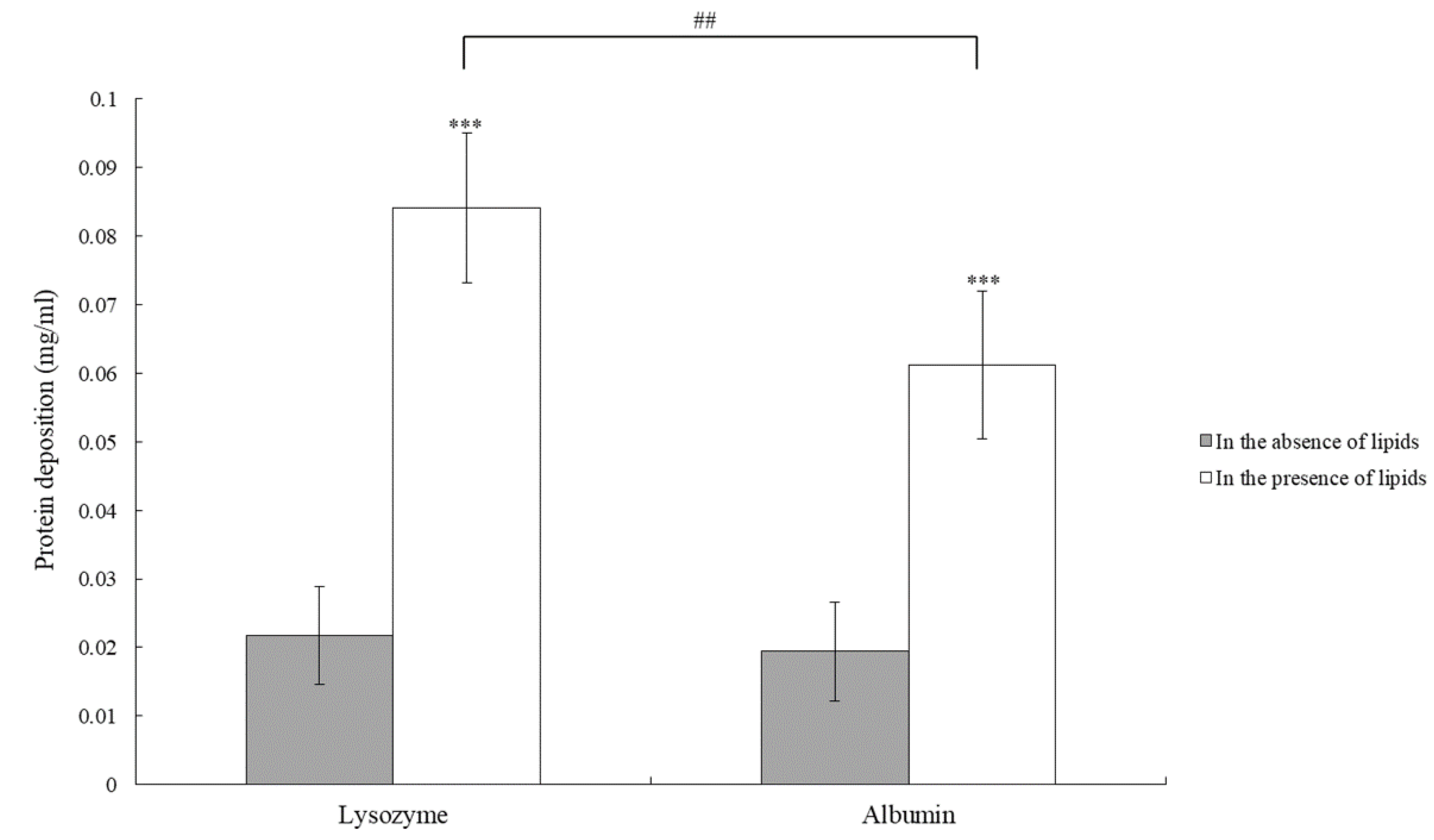
Figure 3. The concentration of protein deposition is accumulated when the lens is not rubbed (white or dark gray confined) or is rubbed (black or calorie-free grayness confined) for cleaning. ** p < 0.01 and *** p < 0.001 when comparing protein deposition amount with lysozyme concentration on the lens without rubbing. && p < 0.01 and &&& p < 0.001 when comparing albumin deposition on no ribbing lens versus lysozyme deposition on rubbing lens. # p < 0.05, ## p < 0.01, and ### p < 0.001 when comparing albumin deposition on the lens without rubbing versus with rubbing. $ p < 0.05 when comparing lysozyme versus albumin degradation on the lens with rubbing. Error confined represented standard departure.
Figure 3. The concentration of protein deposition is accumulated when the lens is not rubbed (white or night gray bars) or is rubbed (black or light grayness bars) for cleaning. ** p < 0.01 and *** p < 0.001 when comparison protein deposition corporeality with lysozyme concentration on the lens without rubbing. && p < 0.01 and &&& p < 0.001 when comparison albumin deposition on no ribbing lens versus lysozyme deposition on rubbing lens. # p < 0.05, ## p < 0.01, and ### p < 0.001 when comparing albumin deposition on the lens without rubbing versus with rubbing. $ p < 0.05 when comparing lysozyme versus albumin deposition on the lens with rubbing. Error bars represented standard departure.
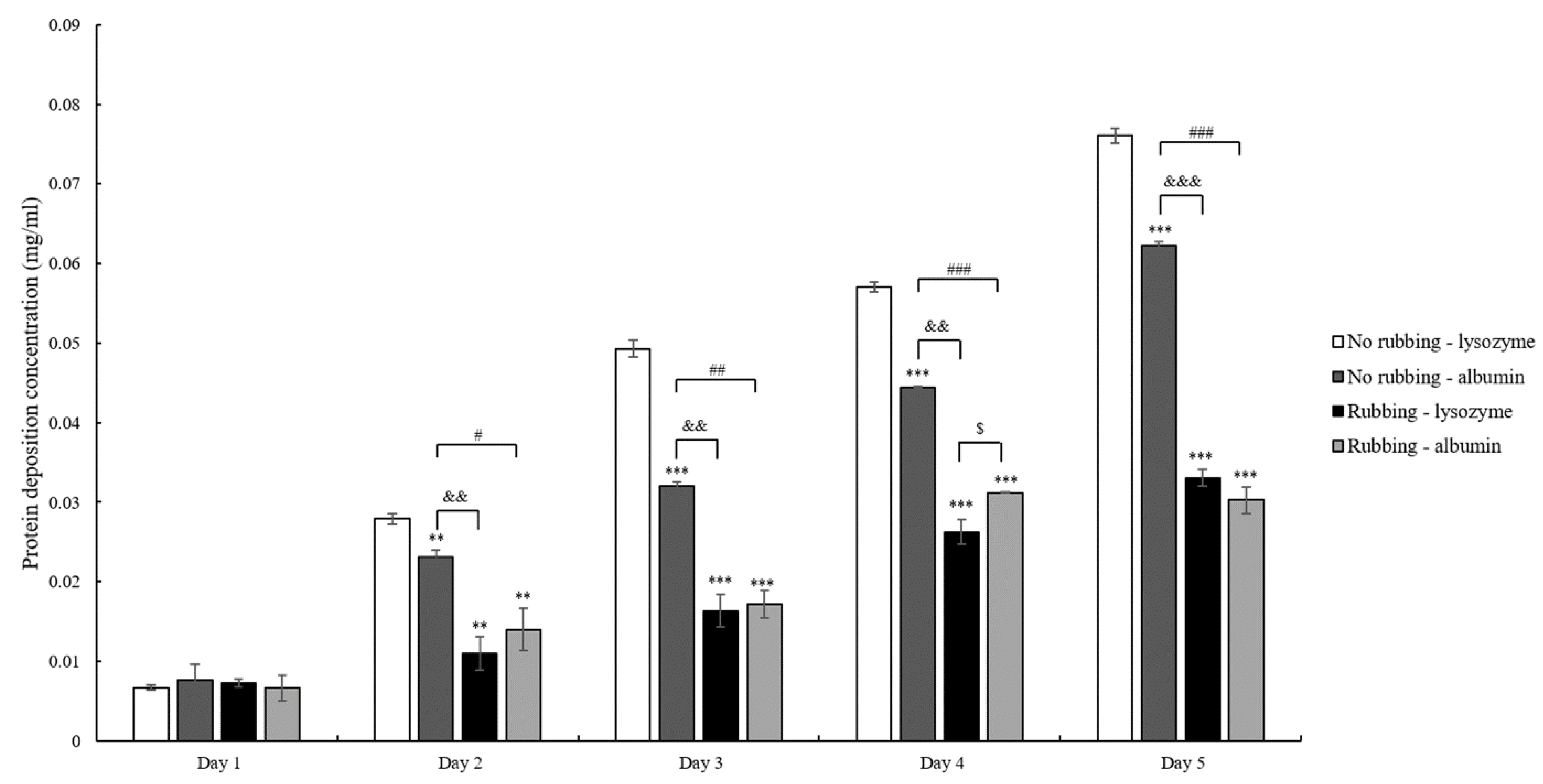
Figure four. Deposited lysozyme concentrations are measured after the lens afterward 1 cycle of procedure. The lens is either not rubbed (gray bars) or rubbed (white bars) at the end of one wheel. The difference is statistically pregnant (p < 0.001) when comparing lysozyme deposition concentration on no rubbing versus rubbing lenses on the aforementioned day. Error confined represented standard deviation.
Effigy 4. Deposited lysozyme concentrations are measured afterwards the lens after one bike of process. The lens is either not rubbed (gray bars) or rubbed (white bars) at the end of one bike. The difference is statistically significant (p < 0.001) when comparing lysozyme degradation concentration on no rubbing versus rubbing lenses on the same day. Fault confined represented standard deviation.
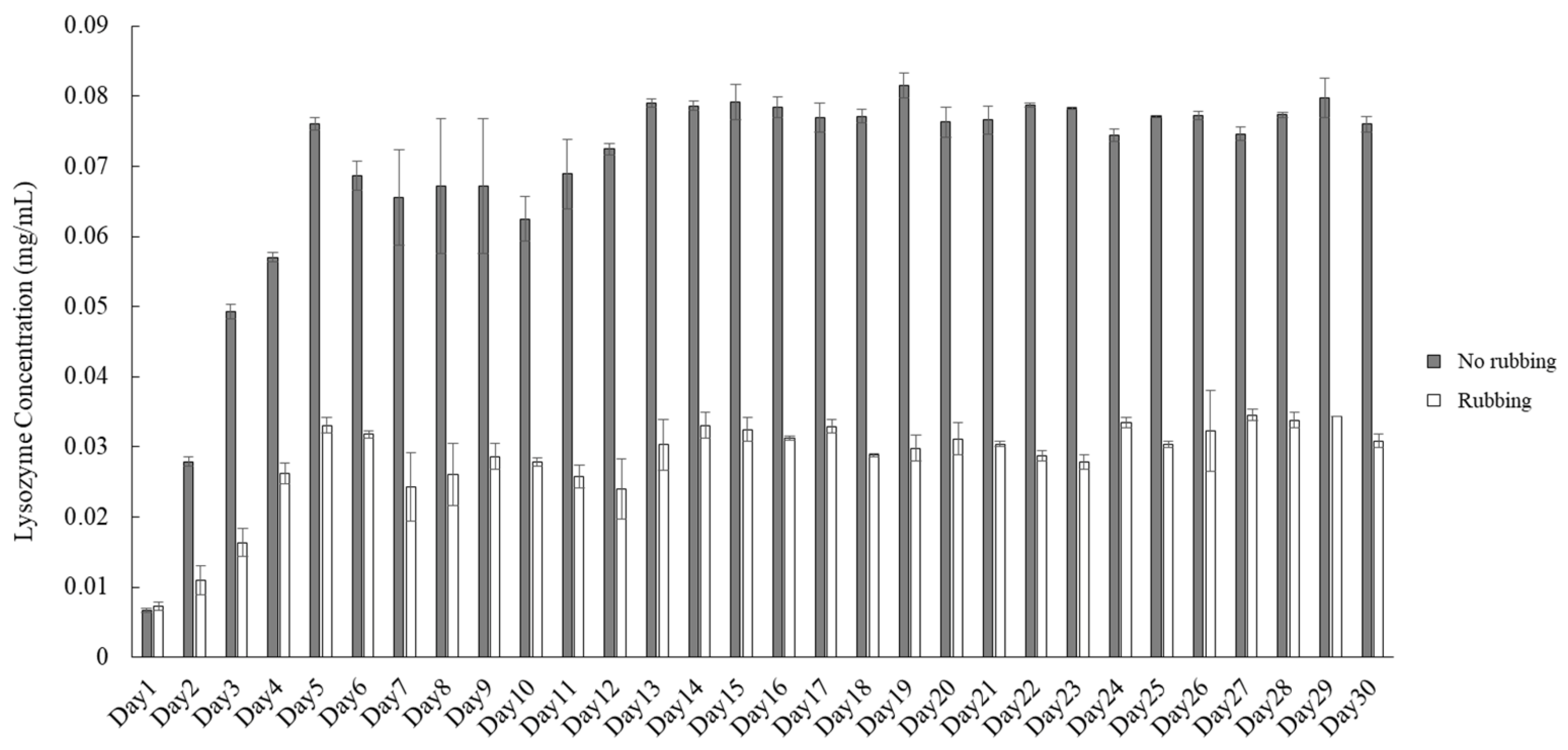
Figure 5. Pictures of lens surface after xxx cycles of poly peptide degradation procedure when the lens was non rubbed (A) or rubbed (B) at the terminate of each cycle.
Effigy five. Pictures of lens surface afterwards 30 cycles of protein degradation procedure when the lens was not rubbed (A) or rubbed (B) at the end of each cycle.

Table 1. The concentration of each component in bogus tear solution.
Table one. The concentration of each component in artificial tear solution.
| Category | Component | Stock Concentration (mg/mL) | Concluding Concentration in Artificial Tear Solution (mg/mL) |
|---|---|---|---|
| Circuitous of salt solution | Sodium chloride | Northward.A. | 5.26 |
| Potassium chloride | 1.19 | ||
| Sodium citrate | 0.44 | ||
| Glucose | 0.036 | ||
| Urea | 0.072 | ||
| Calcium chloride | 0.07 | ||
| Sodium carbonate | ane.27 | ||
| Potassium hydrogen carbonate | 0.xxx | ||
| Sodium phosphate dibasic | 3.41 | ||
| Hydrochloric acid | 0.94 | ||
| ProClin 300 | 200 μL/liter of solution | ||
| Lipid stock solution | Oleic acid | three.6 | 0.0018 |
| Oleic acid methyl ester | 24.0 | 0.012 | |
| Triolein | 32.0 | 0.016 | |
| Cholesterol | 3.6 | 0.0018 | |
| Cholesteryl oleate | 48.0 | 0.024 | |
| Phosphatidylcholine | 1.0 | 0.0005 | |
| Protein | Lysozyme | N.A. | ii.0 |
| Albumin | 0.two |
Table two. Optical specifications of each tested lens earlier and afterwards 30 cycles of protein deposition procedure.
Table 2. Optical specifications of each tested lens earlier and after 30 cycles of poly peptide deposition process.
| Cleaning Method | Item | Before | After | |||||
|---|---|---|---|---|---|---|---|---|
| BC (mm) | CT (mm) | PW (degree) | BC (mm) | CT (mm) | Prisoner of war (degree) | Contact Bending (°) | ||
| No rubbing | 1 | 8.threescore | 0.23 | +0.69 | viii.60 | 0.23 | +0.66 | 15.09 |
| 2 | 8.73 | 0.22 | +0.77 | 8.72 | 0.22 | +0.71 | 23.71 | |
| three | 9.11 | 0.23 | +1.10 | 9.ten | 0.23 | +1.eleven | xiii.02 | |
| Rubbing | ane | 9.01 | 0.24 | +0.93 | viii.92 # | 0.24 | +0.74 | 80.29 |
| 2 | 8.57 | 0.23 | +0.76 | 8.57 | 0.24 | +0.76 | 64.56 | |
| 3 | viii.71 | 0.21 | −0.62 | 8.71 | 0.24 # | −0.48 | 52.32 | |
Table 3. Optical transmission of each tested lens before and after 30 cycles of protein degradation procedure.
Table iii. Optical transmission of each tested lens before and after 30 cycles of protein deposition process.
| Cleaning Method | Item | Before | Later | ||||
|---|---|---|---|---|---|---|---|
| VIS | UVA | UVB | VIS | UVA | UVB | ||
| No Rubbing | i | 83.85 | 12.63 | 0.734 | 85.18 | 12.86 | 0.779 |
| 2 | 88.06 | 13.96 | i.061 | 83.26 | 12.89 | 0.909 | |
| 3 | 82.98 | 12.24 | 0.677 | 76.69 | 11.52 | 0.785 | |
| Rubbing | 1 | 87.86 | 13.55 | 0.831 | 78.nineteen | 10.74 | 0.523 |
| 2 | 84.99 | 12.37 | 0.629 | 83.41 | 12.26 | 0.675 | |
| 3 | 84.12 | thirteen.44 | 0.987 | 83.73 | 13.23 | i.014 | |
| Publisher'south Note: MDPI stays neutral with regard to jurisdictional claims in published maps and institutional affiliations. |
© 2021 by the authors. Licensee MDPI, Basel, Switzerland. This article is an open up admission article distributed under the terms and conditions of the Artistic Commons Attribution (CC BY) license (https://creativecommons.org/licenses/by/iv.0/).

Source: https://www.mdpi.com/2073-4360/13/24/4318/htm
Posted by: jonessincing.blogspot.com
0 Response to "How To Clean Ortho K Lense With Boston Multi Action Solution"
Post a Comment